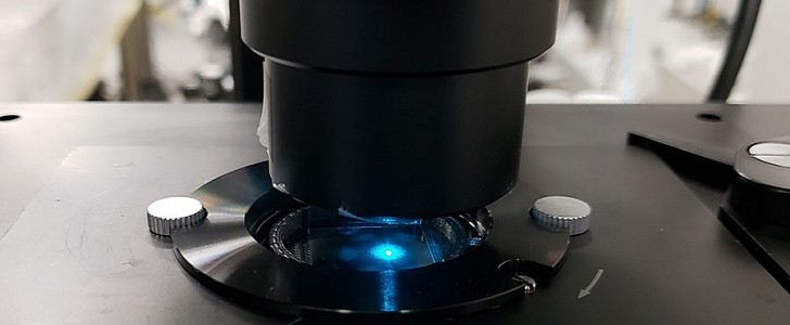
Researchers at the University of California are working on a technology that is able to enhance the resolution of a standard light microscope, allowing it to directly view finer structures and details in living cells.
Usually, light microscopes are used by scientists to see live cells, but they cannot be used to visualize really small things. For example, a regular microscope can have a resolution limit of 200 nanometers (meaning that anything closer than this distance would not be seen as separate objects). The UC engineers' new high-tech microscope slide could provide a much higher resolution limit of 40 nanometers.
According to Zhaowei Liu, a professor of electrical and computer engineering at UC San Diego, finding something that provides a high resolution while also being safe for live cells can be challenging. This new technology aims to do both.
Made up of "nanometers-thin alternating layers of silver and silica glass," the slide can be placed under any microscope. To be able to enhance the image, this thing is coated with a light-shrinking material called a hyperbolic metamaterial.
So how does it work? Scientists explain that the light’s wavelengths that pass through the slide are shortened, and they separate, generating a speckled pattern. This specific pattern will illuminate the sample put on the slide. As a result, low-resolution images of that sample will form. A computer then pieces these images together using reconstruction algorithms to construct a final high-resolution image.
This new tech was already tested by the team of researchers. They were able to clearly tell apart tiny fluorescent beads, and quantum dots spaced 40 to 80 nanometers apart by using a commercial inverted microscope.
Their goal for live cell imaging is to use both high speed and high resolution without damaging the cells when exposed to light. Liu's team is now working to improve the technology so that it can undertake high-resolution imaging in 3D. Currently, their technique creates high-resolution images only in 2D. The researchers' extensive study was recently published in the journal Nature Communications.


